Glaucoma (Green Star)
After cataracts (clouding of the lens), glaucoma is the second most common cause of blindness worldwide. Glaucoma is characterized by progressive damage to the optic nerve, which can lead to blindness if left untreated.
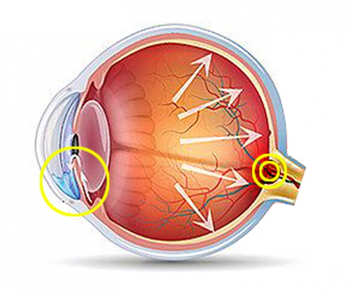
Glaucoma is known as the “silent thief of sight” because it often robs people of their vision without any warning signs or symptoms. In fact, about 2% of people over 40 in Germany have glaucoma, and more than half of those affected are not even aware of it. Glaucoma is a disease that damages the optic nerve. Like a cable, the optic nerve is responsible for transmitting the images we see with our eyes to the brain.
The most common cause of glaucoma is elevated intraocular pressure.
This leads to a slow death of the nerve fibers of the optic nerve. This gradual loss initially leads to an increasing restriction of the lateral visual field. The disease progresses unnoticed in the early stages, as the loss of nerve cells is not associated with symptoms. Without routine eye examinations to check the health of your eyes, glaucoma-related changes may go undetected until the optic nerve is significantly damaged and a large loss of peripheral vision has already occurred. Therefore, regular examinations by an ophthalmologist are important. Left untreated, glaucoma can lead to complete blindness.
Risk factors for the development of who glaucoma
With treatment, glaucoma can often be stopped or at least significantly slowed down. Treatment includes various eye drops to lower intraocular pressure, laser treatments, and surgical procedures.
In recent years, advances in surgical techniques (microinvasive glaucoma surgery or "MIGS") have made glaucoma treatment gentler and safer. These techniques are used in the treatment of glaucoma at the Eye Clinic Regensburg.
Unfortunately, treatment cannot reverse existing damage to the optic nerve.


Optical Coherence Pachymeter
Measurement of Corneal Thickness to Determine Intraocular Pressure Using an OCP (Optical Coherence Pachymeter)
Why is measuring your corneal thickness important?
When screening for glaucoma, your eye doctor measures your intraocular pressure. This measurement is indirect, involving a slight flattening of the cornea using a defined impulse. It assumes an average corneal thickness. However, your actual corneal thickness may vary from this average, which can affect the interpretation of the measurement results.
If your cornea is thinner than average, the measured intraocular pressure will be lower than the actual pressure, potentially causing glaucoma to be overlooked. Conversely, if your cornea is thicker than average, the measured pressure will be higher than the actual pressure.
Using optical pachymetry, your eye doctor can accurately determine your corneal thickness and use this information to calculate your actual intraocular pressure. This is a valuable addition to glaucoma diagnosis, although it is not typically covered by health insurance.
How is corneal thickness measured?
An OCP (Optical Coherence Pachymeter) is used to quickly and accurately measure corneal thickness. This painless procedure does not involve touching your eye, and you can immediately resume your daily activities afterward.
Pentacam
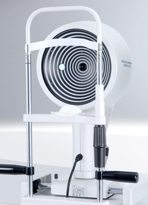
The Pentacam is a specialized camera used for detailed measurements of the front segment of the eye. Using a technique called Scheimpflug imaging, it captures multiple images of the eye within seconds. This provides a comprehensive dataset of 25,000 measurements of height, density, and refractive power in the anterior segment, including the cornea, iris, and lens.
The collected data is stored and can be used for comparison in future examinations to detect even the slightest changes. Upon request, a printout of these measurements can be provided. The procedure is painless and does not require pupil dilation.
In glaucoma, the Pentacam can provide valuable information. In addition to accurately measuring corneal thickness (as with the OCP), it can also measure the angle of the anterior chamber, allowing for an assessment of the structures that drain aqueous humor. This can help determine if lens thickening has contributed to increased intraocular pressure.
Types of Glaucoma
There are different types of glaucoma - not every type of glaucoma is the same or will have the same impact on your life.
This is the most common type of glaucoma. The drainage angle (where the aqueous humor drains from the eye) is open but doesn’t function efficiently enough. This reduced drainage of aqueous humor leads to an increase in pressure inside the eye, which results in a gradual loss of peripheral vision. This can be compared to an air filter that collects dust over time and eventually becomes too clogged to function well
Angle-closure glaucoma occurs when the drainage angle is completely blocked. This type of glaucoma is often found in farsighted eyes of older individuals. It prevents fluid from draining out of the eye, leading to a sudden spike in eye pressure. This extreme pressure increase causes fluid to build up in the cornea, resulting in blurred vision, headaches, severe eye pain, and the appearance of halos around lights.
In this type of glaucoma, the condition is painless and involves a more gradual closure of the drainage angle.
Secondary glaucoma develops when scar tissue, cells, or pigments block the drainage angle. Secondary glaucoma leads to a gradual loss of peripheral vision. Pseudo exfoliative glaucoma and pigment dispersion glaucoma are examples of secondary glaucoma. A secondary glaucoma can also develop from a hemorrhage in the anterior chamber of the eye.
Congenital glaucoma is a rare condition where the drainage angle is impaired or blocked from birth. To prevent blindness, this condition must be treated shortly after birth. Symptoms include enlarged eyes, a cloudy cornea, light sensitivity, and excessive tearing.
Open-angle glaucoma
This is the most common type of glaucoma. The drainage angle (where the aqueous humor drains from the eye) is open but doesn’t function efficiently enough. This reduced drainage of aqueous humor leads to an increase in pressure inside the eye, which results in a gradual loss of peripheral vision. This can be compared to an air filter that collects dust over time and eventually becomes too clogged to function well
Angle-closure glaucoma
Angle-closure glaucoma occurs when the drainage angle is completely blocked. This type of glaucoma is often found in farsighted eyes of older individuals. It prevents fluid from draining out of the eye, leading to a sudden spike in eye pressure. This extreme pressure increase causes fluid to build up in the cornea, resulting in blurred vision, headaches, severe eye pain, and the appearance of halos around lights.
Chronic angle-closure glaucoma
In this type of glaucoma, the condition is painless and involves a more gradual closure of the drainage angle.
Secondary glaucoma
Secondary glaucoma develops when scar tissue, cells, or pigments block the drainage angle. Secondary glaucoma leads to a gradual loss of peripheral vision. Pseudoexfoliative glaucoma and pigment dispersion glaucoma are examples of secondary glaucoma. A secondary glaucoma can also develop from a hemorrhage in the anterior chamber of the eye.
Congenital glaucoma
Congenital glaucoma is a rare condition where the drainage angle is impaired or blocked from birth. To prevent blindness, this condition must be treated shortly after birth. Symptoms include enlarged eyes, a cloudy cornea, light sensitivity, and excessive tearing.
Since many forms of glaucoma are accompanied by an increase in intraocular pressure, a measurement of intraocular pressure in conjunction with the measurement of corneal thickness can provide initial indications of a disease; however, it is not sufficient for a reliable diagnosis. Much earlier, up to six years before the first visual impairments occur, damage to the optic nerve can already be detected with OCT.
To prevent irreparable damage to the optic nerve, please come for regular glaucoma screenings from the age of 40

