Modern Eye Diagnostics
The eyes are our most important sensory organ. Thanks to advances in medicine, many eye diseases can be treated well today. However, a prerequisite for effective therapy is accurate and reliable diagnostics.
At the Eye Clinic Regensburg, we perform complex eye examinations using modern equipment.
The following diagnostic techniques are available to our patients:

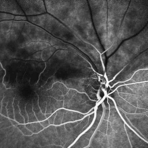
Fluorescein angiography is used to diagnose diseases of the fundus (back of the eye). For this purpose, the vascular system of the retina and choroid is visualized using special dyes (fluorescein, indocyanine green) and captured with a camera.
Fluorescein angiography is used in the diagnosis of age-related macular degeneration, diabetic retinopathy, vascular occlusions, and various inflammatory diseases.
In cataract surgery, the cloudy natural lens is replaced with a clear artificial lens. To achieve optimal surgical results, the eye must be precisely measured before surgery in order to calculate the optimal lens power.
The most modern method today is the highly precise measurement of the eye using a special optical procedure.
In contrast to the older ultrasound method, optical biometry is completely non-contact and is characterized by high precision.
Optical coherence tomography allows for the examination of the individual layers of the retina and the precise localization of changes in the retina. The OCT examination is an important component in the diagnosis of retinal diseases. The findings of the OCT examination are often decisive for the therapeutic decision. By measuring the retinal thickness, the therapeutic success of various therapies can be precisely monitored.
OCT examination has a particular importance in age-related macular degeneration, diabetic retinopathy, vascular occlusions, membranes on the macula (epiretinal gliosis), and macular holes.
Pachymetry is a precise measurement of the corneal thickness. The determination of corneal thickness is an important prerequisite for the interpretation of the measured intraocular pressure in the diagnosis and therapy of glaucoma, as the actual intraocular pressure depends on the corneal thickness. Therefore, pachymetry is performed in the initial diagnosis of glaucoma and in cases of elevated intraocular pressure.
Pachymetry is also used in diagnostics before refractive surgery. Only with sufficient corneal thickness can a laser operation be performed.
Optical coherence tomography (OCT) scans the optic nerve head using a computer-controlled laser system and analyzes it in three dimensions. OCT is used for the early detection of glaucoma. With this examination method, the therapeutic success of glaucoma treatment can be monitored. Changes in the optic nerve can be detected earlier and more clearly with OCT than with other examination methods. Regular examinations provide the ophthalmologist with valuable information for their treatment decisions.
Scanning laser ophthalmoscopy (SLO) is used to examine the retina and choroid of the eye for pathological changes. Using laser light of different wavelengths, the fundus is scanned and examined for pathological changes.
SLO examination plays an important role in the detection of age-related macular degeneration (AMD), diabetic retinopathy, and various other diseases of the fundus and is used in particular for monitoring the course of the disease. Pupil dilation (medical mydriasis) is not necessary for SLO examination.
A Scheimpflug camera allows for a three-dimensional measurement of the anterior segment of the eye. The examination enables the assessment of the anterior and posterior corneal surfaces, the areal determination of corneal thickness, the assessment of anterior chamber depth, and the detection of lens opacities in cataracts.
With the help of modern analysis programs, early detection of various corneal diseases such as keratoconus is possible. The Scheimpflug examination is a routine examination before the surgical correction of refractive errors (“refractive surgery”) and the implantation of special lenses in the treatment of cataracts.
We place great importance on individualized, personal, and compassionate counseling, without losing sight of the patient's overall condition. We are supported by state-of-the-art technology, which allows us to perform a variety of specialized examinations in addition to general eye diagnostics. For the examination of children and neuro-ophthalmological problems, we are supported by an orthoptist.

