Corneal Diseases
About the Cornea
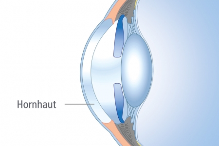
The cornea is the clear, transparent "window" of the eye. The shape of the cornea plays a crucial role in focusing light onto the retina. A normally shaped cornea and a healthy, clear lens allow for sharp light convergence. When the cornea becomes cloudy or irregularly shaped, vision can be impaired. Visiting our corneal specialists can not only improve your vision but also transform your life. Your vision is valuable. The ophthalmologists at the Regensburg Eye Clinic can identify and treat corneal diseases or injuries when wearing glasses or contact lenses no longer meets your needs.
Patients experiencing vision loss due to a diseased or irregularly shaped cornea, such as those with keratoconus, are good candidates for one of many surgical or non-surgical corneal treatments.
Many of the conditions associated with the cornea are genetically inherited from family members, but they can also result from corneal injury or excessive rubbing of the eye. A patient with a diseased cornea may experience distorted vision, light sensitivity, irritation such as a foreign body sensation, blurred vision, swelling, and/or glare. Vision deterioration (including night vision) can often be improved through medical treatment or surgical intervention on the cornea.
Keratokonus
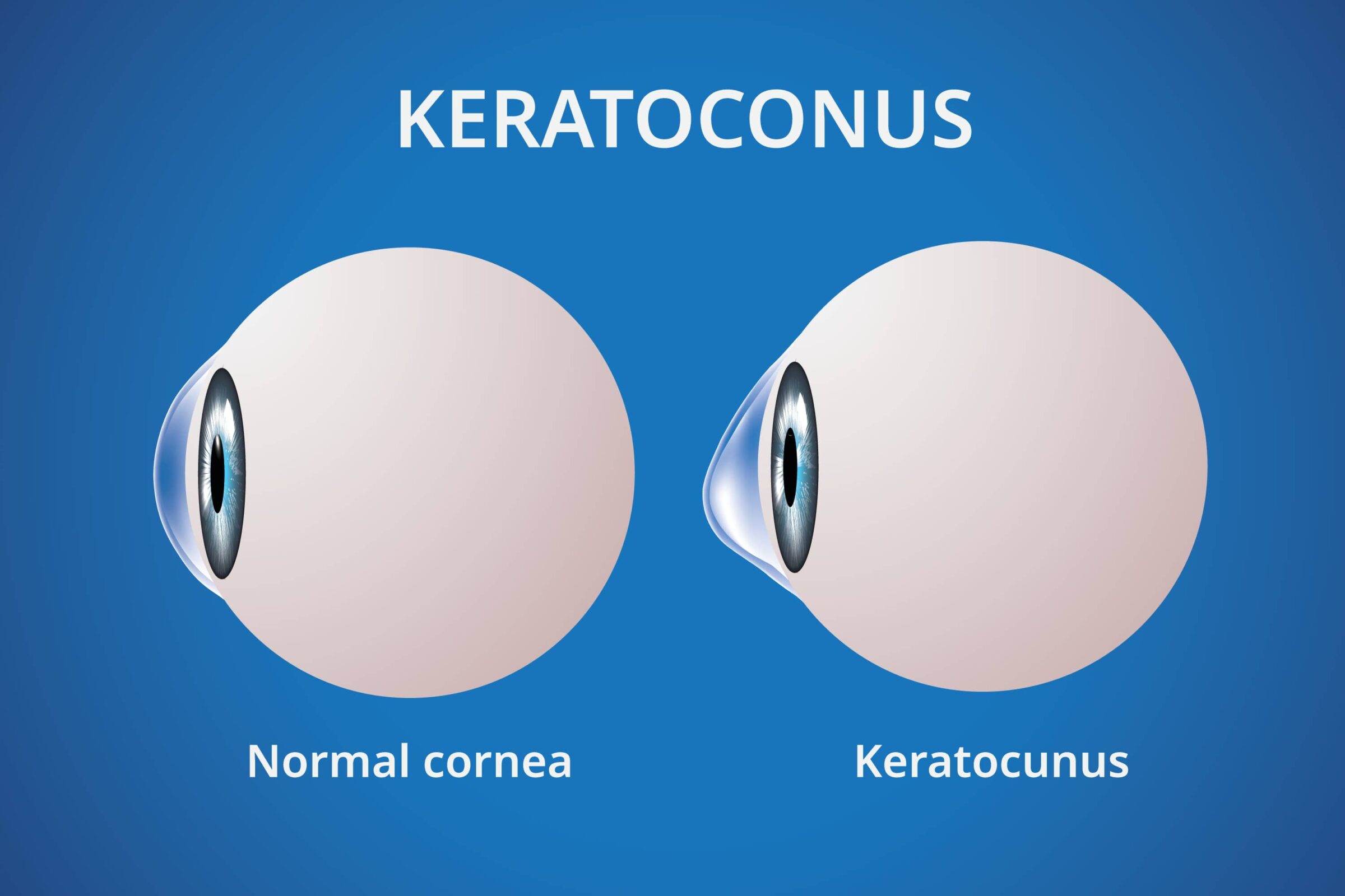
Keratoconus is an eye condition where the cornea becomes thinner and bulges forward in the lower half. A healthy cornea has a round, dome-like shape. In an eye with keratoconus, the cornea develops a cone-like bulge, and this change in shape distorts vision. Keratoconus can make it difficult for individuals to drive, read, and watch television.
The thinning process of the cornea can spontaneously stabilize over a person's lifetime; however, scars may remain on the eye and reduce vision quality. The treatment method for keratoconus depends on the severity of the condition.
In the early stages, eye doctors can manage the condition with glasses or contact lenses. The doctor may use special contact lenses to help artificially smooth the optical surface of the eye, allowing it to refract incoming light more evenly.
As the condition progresses, patients are ideally treated at an early stage with corneal cross-linking. In advanced cases of keratoconus, the cornea may become too thin for cross-linking to be performed. The patient may then require a corneal transplant to replace the damaged cornea with a healthy donor cornea.
Keratoconus is an eye condition where the cornea becomes thinner and bulges forward in the lower half. A healthy cornea has a round, dome-like shape. In an eye with keratoconus, the cornea develops a cone-like bulge, and this change in shape distorts vision. Keratoconus can make it difficult for individuals to drive, read, and watch television.
The thinning process of the cornea can spontaneously stabilize over a person's lifetime; however, scars may remain on the eye and reduce vision quality. The treatment method for keratoconus depends on the severity of the condition. In the early stages, eye doctors can manage the condition with glasses or contact lenses. The doctor may use special contact lenses to help artificially smooth the optical surface of the eye, allowing it to refract incoming light more evenly.
As the condition progresses, patients are ideally treated at an early stage with corneal cross-linking. In advanced cases of keratoconus, the cornea may become too thin for cross-linking to be performed. The patient may then require a corneal transplant to replace the damaged cornea with a healthy donor cornea.

Fuchs’ Corneal Dystrophy
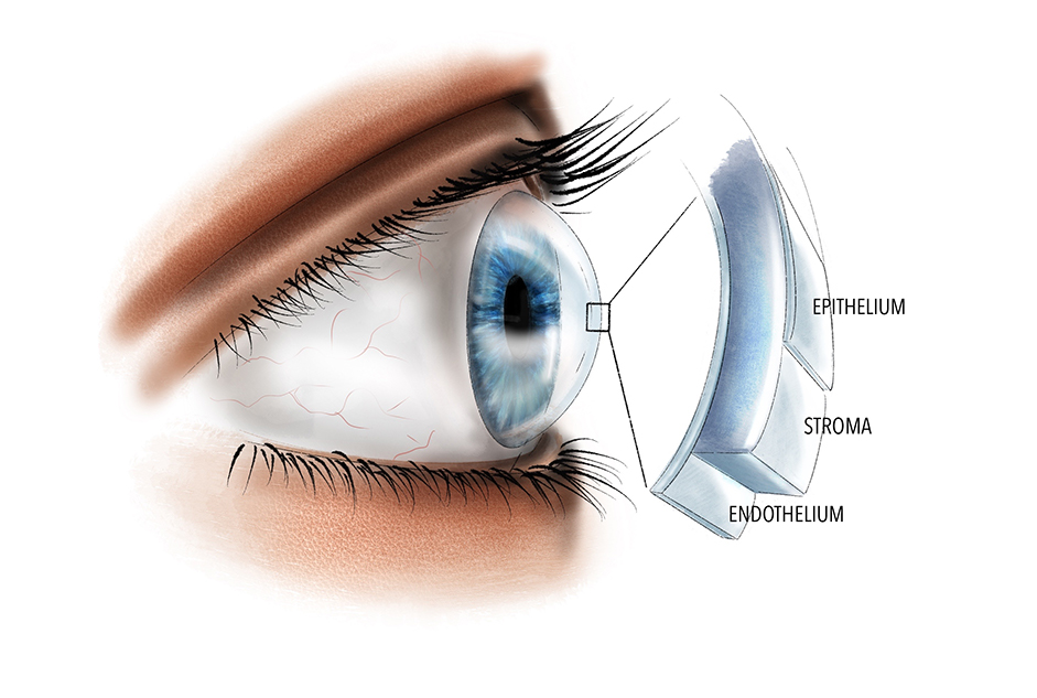
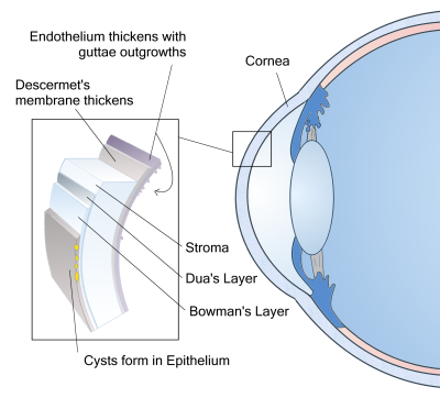
Fuchs’ dystrophy is a genetic condition affecting the cornea. The disease leads to swelling of the cornea because the inner layer of the cornea, the endothelium, progressively deteriorates over a person's lifetime. Endothelial cells regulate the cornea's water balance by pumping out excess fluid. When the number of these cells decreases or their function worsens, fluid begins to accumulate in the cornea. As the cornea swells due to these changes, the patient’s vision deteriorates. In the later stages of the disease, the cornea may develop painful blisters on its surface, a condition known as bullous keratopathy.
Preventive treatment for Fuchs’ dystrophy is not possible. Doctors manage the disease according to its stage. In the early stages, attempts can be made to remove excess fluid from the cornea using specialized decongestant eye drops.
If a patient has elevated intraocular pressure in addition to Fuchs’ dystrophy, glaucoma eye drops can help lower the eye pressure. High eye pressure can contribute to the progression of Fuchs’ dystrophy, so maintaining well-regulated eye pressure is crucial for patients with both conditions.
When Fuchs’ dystrophy has progressed to a point where it significantly impairs the patient’s vision, a corneal transplant can restore vision. An alternative to a full corneal transplant is a transplantation of only the inner endothelial layer of the eye. In these procedures (DMEK/DSAEK), the affected cells are replaced while the outer layer of the eye remains intact, allowing for a faster recovery of vision.
Corneal Transplantation
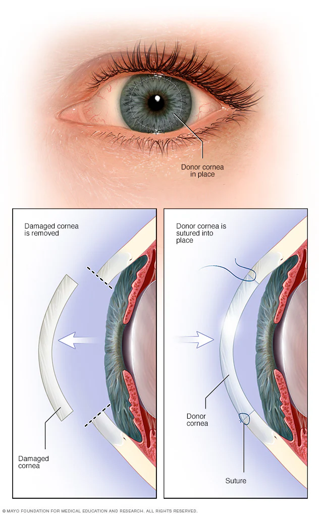
Corneal transplantation is a surgical procedure in which a surgeon replaces a portion of a patient’s diseased cornea with healthy donor tissue, thereby restoring the potential for improved vision. Patients with a cloudy or highly irregular cornea that prevents acceptable vision are candidates for a corneal transplant. This procedure allows patients to achieve clear vision through the complete replacement of the cornea with a donor cornea.
During a corneal transplant, the central part of the cornea is replaced with a donor corneal graft. The surgeon begins by carefully removing the diseased central area of the cornea. A suitable donor tissue is then placed in its position. The surgeon uses stitches to secure the donor tissue to the existing corneal tissue. Corneal transplantation enables patients who have experienced significant vision loss due to a damaged cornea to see better with a clearer and more regular cornea.
