Retinal Detachment
Retinal detachment results in damage to the light-sensitive nerve tissue of the eye. Without treatment, retinal detachment usually leads to blindness. Therefore, symptoms should be taken seriously and evaluated promptly.

What is a Retinal Detachment?
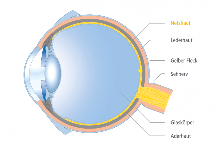
The retina is a thin layer of nerve tissue that lines the inside of the eyeball. A retinal detachment occurs when holes develop in the retina. Through these holes, liquefied vitreous humor can pass beneath the retina. The vitreous humor is the jelly-like substance that fills the back of the eyeball. The fluid beneath the retina causes it to lift away from the retinal pigment epithelium. This layer normally nourishes the inner layers of the retina. Without treatment, a retinal detachment usually leads to blindness in the affected eye.
Most retinal detachments occur as part of the natural aging process of the eye. Changes in the vitreous humor can create traction on the retina, causing it to develop tears or holes. Some people are at increased risk of developing a retinal detachment. These include people who are nearsighted or who have suffered a blunt injury to the eye. In some families, retinal detachments can occur more frequently due to genetic factors.
What do affected people notice?
Affected people often describe light phenomena such as flashes of light. These are perceived more frequently in the dark. Newly occurring streaks or spots that float through the visual field (mouches volantes or floaters) are also frequently described. If a small blood vessel in the retina tears during a retinal tear, a hemorrhage into the vitreous body can lead to completely blurred vision.


Treatment of a Retinal Detachment
In the following, we would like to inform you about the therapy of collagen cross-linking. We would also be happy to provide you with more information about this innovative therapy during a consultation.
Indenting Surgery
In this procedure, a so-called ‘plomb’ or a ‘buckle’ made of silicone material is sewn onto the sclera (the white part of the eye) from the outside. This indents the eyeball in the area of the retinal hole, bringing the retina back into contact with the nourishing and supporting tissue of the retinal pigment epithelium. The silicone material usually remains permanently on the eyeball. During the operation, the underlying retinal hole is treated with a cold treatment (cryopexy). This creates a firm bond between the retina and its underlying tissue.
Vitrectomy
A vitrectomy involves the removal of the altered vitreous body, which is often the cause of a retinal detachment. Today, this is done minimally invasively through three small access points with a diameter of less than 1 mm (25-gauge vitrectomy, keyhole surgery). The retinal holes are then visualized and treated with a laser or cold treatment. This treatment creates a firm bond between the retina and its underlying tissue. This usually occurs after 10 days. During this time, the retina is held in place by a gas bubble or a silicone oil bubble.
High-precision Fine Diagnosis of the Retina
Often, the retina in the back of the eye cannot be adequately assessed with conventional examination methods. Optical coherence tomography (OCT) makes it possible to even assess the internal structures of the multi-layered retina. The measurement is non-contact and takes only a few seconds. By providing a precise visualization of the different retinal layers, OCT examination opens up a new dimension of diagnosis and monitoring for various retinal diseases.

