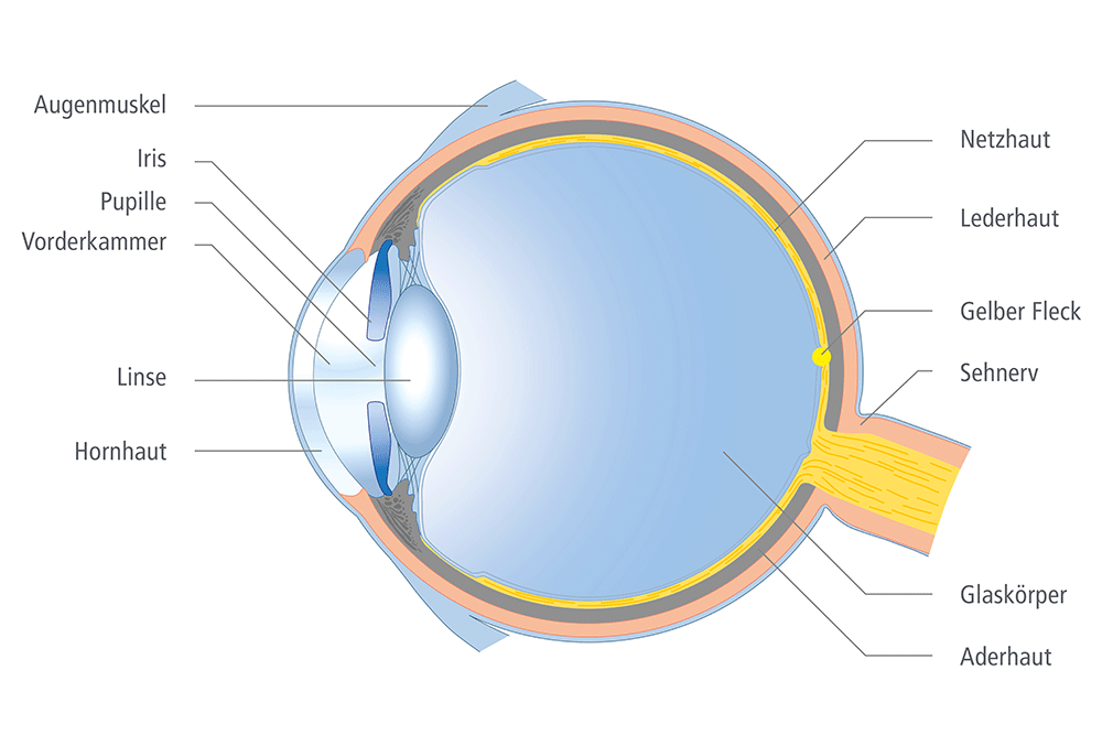
The Structure of the Eye

The structure of the eye is essentially comparable to that of a camera.
The cornea and the lens are similar to the lens system of a camera objective. The iris, together with the pupil, forms the diaphragm: depending on the incident light, the pupil dilates or constricts.
Focusing is achieved by the deformation of the lens. If the gaze is directed at closer objects, for example, the refractive power of the lens increases accordingly. The retina finally corresponds to the film of a camera: here the incident light rays meet and the resulting image is transmitted to the brain via the optic nerve.
Clear vision occurs when the light is bundled by the cornea and lens in such a way that it falls exactly on the yellow spot of the retina (“macula”), the point of sharpest vision.
The Eye from A to Z
Allergic Dermatitis/Conjunctivitis
When the eye is exposed to a substance (allergen) to which it is allergic, it reacts with an inflammatory reaction of the conjunctiva (allergic conjunctivitis) or an inflammation of the skin of the eyelids (allergic dermatitis). Common allergens include pollen, makeup, eye drops, and over-the-counter ointments. Both eyes generally react similarly, but sometimes one eye reacts significantly more. An allergic dermatitis or conjunctivitis can cause symptoms such as redness, eyelid swelling, and watery and crusty eyes.
The eyes can also be irritated by substances even if they do not trigger an allergic reaction. Substances such as soaps, detergents, acids, and solvents can cause people to have the same symptoms as with true allergens.
Treatment options for allergic dermatitis/conjunctivitis include soothing ointments, oral antihistamines, and cool compresses. The only sure cure is to find and avoid the allergenic substance that caused the eye’s reaction. An allergist can help find a long-term solution for allergic dermatitis.
Astigmatism (Curvature of the Cornea)
Astigmatism occurs when an eye has an irregular shape. Imagine a normal eyeball that is shaped like a ball. With astigmatism, the eye is more like an egg shape. This irregularity usually occurs on the surface of the eye, called the cornea. Although astigmatism is generally considered a normal variation and not a disease, it leads to blurred vision without appropriate compensation. With mild astigmatism, the complaints are minor. With higher astigmatism, vision can be corrected by wearing glasses or contact lenses.
Conjunctivitis
Eine Bindehautentzündung tritt auf, wenn die Bindehaut, die den Augapfel umgibt, gereizt wird. Eine Bindehautentzündung kann durch Bakterien, Viren, Allergien oder Chemikalien verursacht werden. Eine bakterielle Konjunktivitis wird im Allgemeinen nicht durch Laborkulturen diagnostiziert, da die Tests teuer und langwierig sind. Zu den Symptomen der Krankheit gehören geschwollene und juckende Lider, ein gelber Ausfluss und kratzende, gerötete Augen. Antibiotische Tropfen und Kompressen lindern in der Regel die Symptome und beenden die Entzündung innerhalb weniger Tage. Bleibt die bakterielle Bindehautentzündung unbehandelt, können schwerwiegende Komplikationen auftreten, wie Infektionen der Hornhaut, der Lider und der Tränenkanäle. Bakterielle und virale Bindehautentzündungen sind hoch ansteckend. Einer Ansteckung kann vorgebeugt werden durch gutes Händewaschen, die Verwendung sauberer Handtücher und das Fernhalten von Menschen, die mit der Krankheit infiziert sind.
Basal Cell Carcinoma of the Eyelid (Basalioma)
Basal cell carcinoma is the most common type of skin cancer on the eyelid, and the tumors are often confused with benign growths. Basal cell carcinoma appears as a pearly or waxy nodule with a central, ulcerated area that does not heal. Excessive sun exposure plays a significant role in the development of basal cell carcinomas. While basal cell carcinomas generally do not spread widely within the body, they can be locally invasive and should be removed promptly. Many patients with a basal cell carcinoma develop another tumor after diagnosis, so they should be regularly examined both at the original site and at other sun-exposed areas.
Blepharitis (Lid Margin Inflammation)
Blepharitis occurs when the tiny oil glands (meibomian glands) near the base of the eyelashes become blocked. This leads to irritated, red eyes. Although the condition can be painful, it generally does not cause permanent eye damage. Symptoms of blepharitis include watery, red eyes, red eyelids, a burning sensation in the eyes, greasy, itchy or sticky eyelids, scaly skin around the eyes, and crusted eyelashes upon waking. Additional symptoms include excessive blinking, light sensitivity, eyelashes that grow in an abnormal direction, and loss of eyelashes.
Regular eyelid hygiene is used to treat blepharitis. The eyelids are cleaned one to two times a day with warm, moist compresses. If symptoms are not relieved by eyelid care and moisturizing eye drops, a medication prescribed by a doctor can reduce inflammation.
Eyelid surgery
A blepharoplasty is a surgical procedure that can be performed on the upper and lower eyelids to remove excess skin and tighten the tissues surrounding the eye. When the upper eyelid droops, it can cover the pupil and impair the patient’s vision. During a blepharoplasty, the ophthalmologist removes the excess skin on the eyelid so that the eyelid no longer obstructs vision. The procedure can lead to improved vision as well as a more alert appearance of the eyes.
Gray cataract
A cataract is a clouding of the eye’s natural lens. Cataracts develop over time as a person ages. The clouding of the lens reduces the amount of light that can pass through the lens to the retina. Cataracts cause a painless, gradual loss of vision. Cataracts can significantly impair vision. Other symptoms of cataracts include increased sensitivity to glare, poor night vision, and faded colors. If a person’s ability to read or drive is affected or their daily activities are impaired by vision problems, cataract surgery is the best solution.
Chalazion
A chalazion is a blockage of an oil gland in the eyelid. Chalazia can vary in size, and can grow as large as a pea. In the initial stages, chalazia are often painful and tender. Over time, they become firm and painless. Without treatment, astigmatism and blurred vision can result. Chalazia can sometimes disappear on their own over time. Treatment for chalazia consists of warm compresses, eye drops and ointments, and oral medications. A surgical procedure can be performed by an ophthalmologist to remove chalazia.
Corneal abrasion
Erosions describe a defect in the superficial covering layer of the eye, the so-called epithelium. Corneal abrasions can be caused by, for example, a branch or a fingernail. They are painful and lead to temporary blurred vision and redness of the conjunctiva. Erosions often heal quickly and without complications. An ophthalmologist can prescribe bandage contact lenses, eye ointments, or pain relievers for treatment. It is important that patients do not rub their eyes vigorously while recovering from a corneal erosion, as it takes time for the new cells to bond with the underlying tissue.
In some cases, there are recurring epithelial defects, referred to as recurrent erosion. This happens when the epithelial cells no longer interlock well with the underlying corneal tissue. Painful erosions then often occur upon waking up in the morning. To treat recurrent erosions, a laser treatment (PTK) is often performed.
Diabetic Retinopathy (Diabetes of the Retina)
Diabetes can lead to damage to the blood vessels in the eye. These damaged vessels can leak fluid or blood, causing the retina to swell. Deposits form at the back of the eye, called hard exudates. Diabetic retinopathy can take on various forms. Background retinopathy describes an early form of the disease where there is no change in vision.
In later stages, serious changes can develop, such as macular edema with a decrease in visual acuity, new blood vessel formation (proliferative retinopathy), and retinal detachment. Symptoms of diabetic retinopathy can include fluctuating vision, blurred vision, or vision loss. In some cases, diabetic retinopathy remains asymptomatic at first. Regular eye examinations with pupil dilation allow the ophthalmologist to diagnose diabetic retinopathy. Through early diagnosis and treatment, vision loss can possibly be prevented.
In some cases, there are recurring epithelial defects, referred to as recurrent erosion. This happens when the epithelial cells no longer interlock well with the underlying corneal tissue. Painful erosions then often occur upon waking up in the morning. To treat recurrent erosions, a laser treatment (PTK) is often performed.
Ectropion and Entropion
When an eyelid loses its tone, the eyelid margin can turn inward (entropion) or outward (ectropion). Ectropion generally occurs in the lower eyelid of older people. The eyelid margin turns away from the eye and the conjunctiva on the inside of the lower eyelid is exposed. The eye tears and is irritated. Entropion describes a condition where the eyelid turns inward toward the surface of the eye. The cause is usually an imbalance in the eyelid muscles, which occurs with increasing age. The eyelashes rub against the cornea and the eye tears. Another possible cause of an eyelid malposition is a lid scar after an injury. Treatment generally consists of a surgical procedure.
In later stages, serious changes can develop, such as macular edema with a decrease in visual acuity, new blood vessel formation (proliferative retinopathy), and retinal detachment. Symptoms of diabetic retinopathy can include fluctuating vision, blurred vision, or vision loss. In some cases, diabetic retinopathy remains asymptomatic at first. Regular eye examinations with pupil dilation allow the ophthalmologist to diagnose diabetic retinopathy. Through early diagnosis and treatment, vision loss can possibly be prevented.
In some cases, there are recurring epithelial defects, referred to as recurrent erosion. This happens when the epithelial cells no longer interlock well with the underlying corneal tissue. Painful erosions then often occur upon waking up in the morning. To treat recurrent erosions, a laser treatment (PTK) is often performed.
Glaucoma
Glaucoma is the second leading cause of blindness worldwide. Over time, the disease damages the nerve fibers of the optic nerve. Glaucoma can cause visual field defects, and eventually all fibers of the optic nerve can be destroyed. This can result in complete blindness. Early diagnosis and treatment of the disease can limit damage to the optic nerve and prevent resulting blindness.
In chronic open-angle glaucoma, which typically occurs after age 45, the drainage system of the eye ages and weakens. This results in an increase in intraocular pressure. Another form of glaucoma, called angle-closure glaucoma, occurs when the drainage system is completely blocked, leading to sudden blurred vision, eye pain, headache, halos around lights, and nausea. Although glaucoma cannot be reversed, its effects can be slowed by lowering intraocular pressure.
In later stages, serious changes can develop, such as macular edema with a decrease in visual acuity, new blood vessel formation (proliferative retinopathy), and retinal detachment. Symptoms of diabetic retinopathy can include fluctuating vision, blurred vision, or vision loss. In some cases, diabetic retinopathy remains asymptomatic at first. Regular eye examinations with pupil dilation allow the ophthalmologist to diagnose diabetic retinopathy. Through early diagnosis and treatment, vision loss can possibly be prevented.
In some cases, there are recurring epithelial defects, referred to as recurrent erosion. This happens when the epithelial cells no longer interlock well with the underlying corneal tissue. Painful erosions then often occur upon waking up in the morning. To treat recurrent erosions, a laser treatment (PTK) is often performed.
Herpes Simplex (Eye)
When the herpes simplex virus type I infects the eye, it can impair vision and lead to corneal ulcers, eyelid blisters, and eye inflammation. In many cases, the virus can return after successful treatment. A herpes infection of the eye can cause eye redness, tearing, light sensitivity, and a foreign body sensation. In a chronic course, it can lead to permanent inflammation and scarring of the cornea. Treatment to kill the virus is essential to prevent scarring and permanent vision loss.
Hyperopia (Farsightedness)
Hyperopia, or farsightedness, occurs when the eyeball is too short relative to the refractive power of the cornea and lens. Due to the shorter distance between the front of the eye, the cornea, and the retina at the back of the eye, the light rays focus behind the retina. With hyperopia, distant objects appear clear, while nearby objects appear blurry.
Iritis (Uveitis)
Iritis describes an inflammation of the iris, the colored part of the eye. In many cases, iritis can be linked to a variety of diseases or infections. However, in some cases, no clear cause can be found (idiopathic iritis). Symptoms of iritis include red eyes, light sensitivity, blurred vision, tearing, or eye pain. Iritis is often a recurring problem that requires repeated treatment with medications and eye drops to reduce inflammation. Complications such as cataracts, glaucoma, and corneal changes can occur due to iritis and its treatment. Therefore, close monitoring by an ophthalmologist is essential for effective management.
Migraine (Eye)
When the blood vessels in the head constrict rapidly and restrict blood flow to the brain, it can lead to migraine headaches and visual disturbances. A restriction of vision in part of the visual field or seeing flickering lights may be a person’s only symptoms (ocular migraine). Headaches and nausea associated with migraines are largely caused by a reflexive widening of the blood vessels.
Myopia (Nearsightedness)
If the eye is too long in relation to the refractive power of the cornea and lens, this is referred to as nearsightedness (myopia). Due to the greater distance between the surface of the eye and the retina at the back of the eye, the light rays focus in front of the retina. With nearsightedness, near objects appear sharp, while distant objects appear blurry.
Retinal Detachment
A retinal detachment occurs when the innermost layer of the eye (the retina) separates from the underlying choroid. A retinal detachment can happen when the jelly-like substance inside the eye (vitreous humor) begins to liquefy and change. When this occurs, affected individuals may see floaters in their vision. The vitreous humor can pull on the retina, causing flashes of light. This pulling can sometimes create a hole in the retina, allowing fluid to pass underneath and detach the retina. If holes in the retina are found early, they can be sealed with laser treatment. If a retinal detachment has already occurred, surgery is necessary.
Presbyopia
Presbyopia describes a natural aging process of the eye. Inside a healthy eye, the lens changes its shape depending on the distance of the object it is trying to see. When an object is close to the person, the lens becomes rounder, and when an object is far away, the lens becomes thinner.
Over time, the eye loses some of its ability to change shape, leading to presbyopia. From the age of 40, people notice a blurriness when they try to focus on nearby objects.
If patients are nearsighted, they can still see clearly up close without glasses, despite having presbyopia.
Pterygium (Winged Flesh)
A pterygium is a growth of the conjunctiva that can occur on the surface of the cornea, especially if a person has been frequently exposed to the sun, wind, dust, and other harsh climatic conditions. Pterygia are more common in men than in women. Pterygia can grow larger over time or remain stable and unproblematic for a long time. Symptoms include blurred vision, dry eyes and irritation, as well as redness during growth. Good protection from ultraviolet radiation can prevent the development of pterygia. Eye drops can help with the symptoms of dry eyes. Surgical removal can be performed for visual or cosmetic reasons.Over time, the eye loses some of its ability to change shape, leading to presbyopia. From the age of 40, people notice a blurriness when they try to focus on nearby objects.
If patients are nearsighted, they can still see clearly up close without glasses, despite having presbyopia.
Vitamins
For most people in Europe, a balanced diet provides their eyes with sufficient vitamins. Although cataracts are associated with malnutrition in developing countries, the development of cataracts due to a lack of essential vitamins is not considered a problem in Germany.
In older people, taking a multivitamin supplement with vitamins E, C, and beta-carotene, as well as the minerals zinc and selenium, can help reduce the risk of developing macular degeneration.If patients are nearsighted, they can still see clearly up close without glasses, despite having presbyopia.
Central Retinal Vein Occlusion
The central retinal vein drains all the blood from the retina into the bloodstream. When the central retinal vein becomes blocked, it often causes a sudden loss of vision. In some cases, vision may improve over time, while in others, vision continues to deteriorate. Risk factors for developing a central retinal vein occlusion include diabetes and high blood pressure.In older people, taking a multivitamin supplement with vitamins E, C, and beta-carotene, as well as the minerals zinc and selenium, can help reduce the risk of developing macular degeneration.If patients are nearsighted, they can still see clearly up close without glasses, despite having presbyopia.

